

a knot in your stomach. When undergoing a urinary tract surgery, it is completely understandable that you will experience anxiety. You may be asking yourself, what does this test entail? Does it hurt? What will the results tell me?
I am Dr. Sandeep Nunia, Head Urology Consultant expert in reconstructive urology. At Manav Urology Hospital in Jaipur, we recognize the trauma that patients encounter during these specialized tests. This guide will ensure that you know all the details about RGU MCU urethrography so you don’t enter the appointment at our Urology hospital jaipur with a jittery feeling.
1. What is Urethrography? RGU and MCU Explained
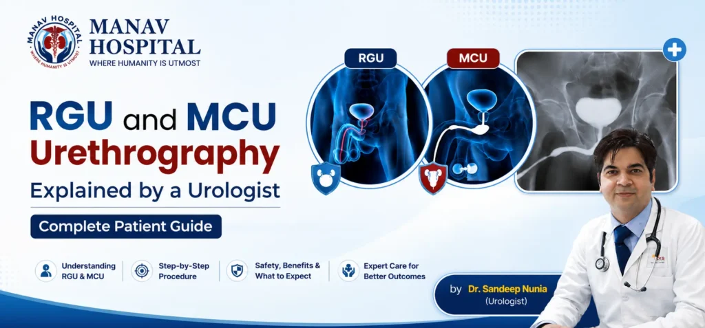
Urethrography is a specialized type of X-ray imaging technique that helps to specifically visualize the structure and integrity of the tube that carries urine from the bladder out of the body (urethra). This is because the fine contours of this delicate pathway can not be easily mapped with a standard ultrasound or a CT scan, so we run an RGU test or an MCU test for them.
The medical terms help, if you consider the urinary tract like a one-way street:
- Retrograde Urethrography also called ascending urethrography (ASU) is an image that examines the urethra from outside in. A liquid contrast dye is carefully instilled into the urinary meatus (meatoscopic) and then pushed back into the system, against the urine’s normal flow, to outline any constriction or blockage.
- Also called voiding cystourethrography (VCUG) or descending urethrography, this is a view of the system in reverse. Your anatomy is then seen on X-rays as you pass the contrast dye into the bladder through the temporary catheter and then pass the dye out as you urinate (micturate) it out of your bladder.
To summarise: RGU considers the front part of the passage when relaxed, while MCU assesses the bladder neck and the internal structures as they are being emptied.
2. Why Your Urologist Recommended This Test
These advanced imaging tests help to identify structural problems that may be responsible for obstructive urinary symptoms, recurrent infection or pelvic pain.
Key Indications for RGU
The most common reason for the test is for urethral strictures. It refers to the abnormal narrowing of the lumen (the inside channel) of the urethra. Gonorrhoeal urethral stricture infection or non-specific urethral infection in the past is a source of severe urethral strictures in India.
- Post-Catheterisation Injury: It is a very common phenomenon in all clinical conditions of Indian and in the surrounding world. A urinary catheter that is stuck in place or inserted incorrectly may cause scar tissue to develop which over time can obstruct natural urination.
- Urethral Trauma: Complex urethral stricture diagnosis parameters or structural tears after road traffic accidents and pelvic fractures.
- Anatomical Defects: Detection of a urethral fistula (abnormal tunnel bypass), diverticulum (outpouching) or suspected lower urinary tract malignancies.
Key Indications for MCU
- Vesicoureteral Reflux (VUR): This is the gold standard test in MCU for child India clinical protocols. It tests for any urine backup from the bladder to the kidneys that occurs abnormally and that may lead to permanent kidney damage.
- Posterior Urethral Valves (PUV): Congenital (birth) defect in newborn boys that occurs due to the presence of abnormal tissue flaps obstructing the urine passage from the bladder.
- Neurogenic Bladder & Obstructions: Assessing bladder outlet obstructions, pelvic floor dyssynergia, or bladder dysfunction caused by spinal and neurological conditions.
The word RGU or MCU does not indicate a dangerous condition of your body. These are standard, very effective diagnostic maps. The entire session takes about 20-30 minutes and most patients proceed through it completely.
3. What is the Difference Between RGU and MCU?
Comparing these dynamic investigations is easy with the two below reference tables that outline the workings of RGU vs MCU urethrography, and other materials used in urology such as RGU vs cystoscopy.
RGU vs MCU Core Comparison
| Feature | RGU (Retrograde Urethrography) | MCU (Micturating Cystourethrogram) |
| Full Form | Retrograde / Ascending Urethrography | Micturating / Voiding Cystourethrogram |
| Primary Purpose | Image the anterior (front) urethra | Image the posterior urethra and bladder during voiding |
| Contrast Direction | Inserted via urethral tip (retrograde flow) | Instilled into bladder via catheter, then voided out |
| Part Examined | Penile and bulbar urethra primarily | Bladder neck, posterior urethra, and ureters (for reflux) |
| Patient State | At rest — no active urination required | During active urination (voiding phase) |
| Common Patient Mix | Primarily adult males | Adults, young boys, and paediatric female children |
| Key Diagnostics | Urethral strictures, trauma, fistulas | VUR grading, posterior urethral valves, incontinence |
| Average Duration | 15–20 minutes | 20–30 minutes |
Urethrography vs Alternative Urological Tests
| Feature | RGU / MCU Urethrography | Cystoscopy | Urinary Ultrasound |
| Invasiveness | Minimally invasive | Invasive (rigid or flexible camera scope inserted) | Completely non-invasive |
| Anatomical Data | Accurate stricture length, location, and backflow | Direct visual view of the inner tissue lining and walls | Static volume of bladder, prostate size, or kidney stones |
| Clinical Strength | Ideal for stricture surgery planning & VUR | Best for finding bladder tumours or small stones | Best for baseline tissue checks |
4. How RGU is Performed: Step-by-Step
Knowing exactly what the steps are in your RGU means that you don’t have to worry about the unknown. Let’s take a look at what your RGU experience will be like from your perspective as a patient:
- Positioning: You will be lying down on a comfortable X-ray table with your back facing the X-rays. You will then be asked to bend a leg and lean slightly to one side to get the pelvis at an optimum angle.
- Cleansing: The medical staff completely cleanses the tip of the penis using a cold, sterile antiseptic solution to ensure that no bacteria enter the penis.
- The placement of the catheter: The catheter tip is a small, soft one or a special syringe nozzle is gently placed at the very opening of the urethra. This is not a normal urinary catheter and will not be inserted far down into your bladder.
- Contrast Injection: A dye dye used to make the X-rays easier to see is injected slowly through the opening. The tube is gently inflated with this fluid in order to be captured in the X-ray at its natural shape.
- The real-time images are taken with digital fluoroscopy equipment, and our specially-trained radiologist makes 2-3 images. May need to hold breath for a few short seconds.
- Completion: The small end is detached, you dry off any excess liquid, and then you can put your clothes back on.
Is the RGU Test Painful?
The majority of patients experience a pinching pressure in the tip or a warm, full sensation when the contrast liquid is injected, as it enlarges the passage. The first catheter position only takes about 30 seconds to align. It will be a bit uncomfortable or awkward, but not really painful when done by an experienced team.
Here we are using the latest digital fluoroscopy equipment as opposed to the old fashioned conventional X-rays. This can result in a much lower radiation dose, plus high-definition real-time video loops, and shorter time sitting on the table.
5. How MCU is Performed & Paediatric Section
The how is the MCU performed workflow:
- Bladder filling: A very thin and lubricated catheter is inserted into the urethra and then directly into the bladder. A sterile contrast fluid (which contains iodine) is slowly introduced into the bladder until there is a clear, strong sense of needing to go to the toilet.
- Catheter Removal: Temporary tube will be removed cleanly once the bladder is full.
- Voiding Under X-Ray: A urine receptacle will be provided and you will be asked to void your urine naturally, standing or lying tilted on the fluoroscopy table.
- Dynamic captures: During urination, continuous real-time X-rays show the action of the bladder neck opening, and whether fluid is abnormally rising back toward the kidneys.
- Post-Void Check: Final quick check is taken immediately after voiding to get the post-void residual urine volume.
Paediatric VCUG / MCU in Children
For young children this test may be a daunting experience for parents as they are diagnosed. We ensure your child a safe and calming environment by:
- Parental Presence: One parent stays very close by in the room wearing a protective lead apron to maintain the child’s calm and stillness.
- Specially designed equipment: We use ultra soft micro sized paediatric catheters to minimise physical discomfort and we employ strict weight optimisation to reduce radiation.
- No Sedation Needed: This procedure is 20-30 minutes. It is not sedated, since it’s based on the child’s natural need to urinate. Still the best worldwide for Vesicoureteral Reflux Grading and Kidney Protection in developing kidneys.
6. How to Prepare for RGU/MCU: Patient Guide
When you’re properly prepared you’ll be able to get clean images the first time and enjoy reduced risk of the occasional post-procedure side effect.
Before the Test
- Fasting: No fasting is required. Light meals or normal meals may be consumed prior to entry.
- Hydration: As long as there is an MCU, drink plenty of water (3-4 glasses). Well hydrated system makes filling the bladder more comfortable.
- Medical Disclosures: Your urologist should be notified of any known history of contrast dye allergy or iodine sensitivity, blood thinners and/or metformin medications and if you think you have an active UTI.
- Clothing: Two-piece swimsuit for easy changing.
After the Test
- Hydration Plan: Upset water consumption by 2-3 liters for 24 hours. This quickly eliminates any remaining contrast material, and reduces the amount of minor local irritation.
- Normal Sensations: The burning sensation and a pinkish color in the first 1 or 2 urinations is perfectly normal.
- When to call us: If you suddenly get fever or chills or cannot pass urine at all, or you see big blood clots in your urine, call our clinic right away.
7. Understanding Your RGU/MCU Results
After your films are processed by the radiologist, your urologist will look at the “definitive diagnostic structural map”.
Normal vs Abnormal findings.
- Normal report: No narrowing of the urethral outline is seen (no narrowing is seen on the X-ray), no structural narrowing of the urethra is seen, it is smooth and wide and the contrast dye goes nicely into the bladder.
- Urethral Stricture on RGU: Is a distinct constriction or blockage point of the white contrast line. Your urologist will take into account whether the exact length (1 cm vs 3 cm) and location (bulbar, penile, or membranous) will aid in determining the next surgical steps, including an endoscopic visual internal urethrotomy (VIU) or a formal urethroplasty reconstruction.
- Filling Defects: Dark areas within the white contrast track (pathway) that indicate a possible embedded urethral stone, blood clot or a small polyp/tumour.
- Extravasation: The presence of contrast fluid outside the urethral wall, which is associated with a recent traumatic rupture, or a long fistula tract.
- On MCU: Vesicoureteral Reflux (VUR) – contrast refluxing up the ureters and kidneys. A report with a reflux grade of 3 implies that there is severe reflux with the contrast moving up and a minor dilation of the renal pelvic. A medical management or minimally invasive endoscopic correction is warranted in this case.
8. Risks, Complications & Contraindications
These processes are very safe if performed with contemporary clinical practice guidelines, but one must be candid about risks.
- Any instrument has a small risk of carrying up the urinary tract with it (Urinary Tract Infection or UTI). This is overcome at our Clinic with the use of exclusively autoclaved instruments, single-use sterile consumables and a short course of 3 days of preventative oral antibiotics.
- Transient Haematuria: Occurs for short periods of time, but is not very much blood in the urine.
- An uncommon problem that occurs when a catheter passes through a very weak and diseased stricture wall. This risk is minimized by avoiding force and by having a trained urologist or experienced technician do complex cases.
- Radiation Exposure: Diagnostic exposure is very minimal and is similar to a standard pelvic X-ray.
Key Contraindications
If you have any of the following, we will make sure to postpone or reschedule your procedure:
- Active UTI: The procedure should be delayed until a repeat urine culture is negative to avoid transfer of the bacteria to the kidneys.
- Pregnancy: Alternative imaging methods are encouraged because of the risk of radiation to the developing fetus.
- Iodine Allergy (Severe): Rescheduled unless cleared by an immunologist with a protocol for pre-medication.
9. Cost of RGU and MCU Test in India
Price for diagnostics varies from a city to another, as per the tier, facility infrastructure, and type of technological imaging system deployed in a specific facility.
Estimated Diagnostic Cost Breakdown
| Test Type & Facility Profile | Typical Indian Cost Range (INR) |
| RGU / MCU (Standalone Diagnostic Centre) | ₹1,500 – ₹3,500 |
| RGU / MCU (Private Hospital with Digital Fluoroscopy) | ₹4,000 – ₹8,000 |
Factors Dictating Cost
- Technology Choice: New digital continuous fluoroscopy machine is more expensive than the old fixed X-ray film, but it can deliver lower radiation doses, and much clearer images of the patient.
- Contrast, which is made from high quality non-ionic contrast material, significantly decreases the chances of allergic reaction from the contrast, and costs a little more than older ionic contrast materials.
- Insurance & TPA Coverage: They are required tests and not cosmetic treatments, thus the cost of these procedures are covered by most major private health insurance networks, corporate panels and public health insurance schemes (CGHS), as long as they have been accurately prescribed by a registered urologist. Please confirm details of your specific Policy with our Hospital’s billing department prior to admission.Technology Choice: New digital continuous fluoroscopy machine is more expensive than the old fixed X-ray film, but it can deliver lower radiation doses, and much clearer images of the patient.
- Contrast, which is made from high quality non-ionic contrast material, significantly decreases the chances of allergic reaction from the contrast, and costs a little more than older ionic contrast materials.
- Insurance & TPA Coverage: They are required tests and not cosmetic treatments, thus the cost of these procedures are covered by most major private health insurance networks, corporate panels and public health insurance schemes (CGHS), as long as they have been accurately prescribed by a registered urologist. Please confirm details of your specific Policy with our Hospital’s billing department prior to admission.
Taking an RGU or MCU test can be stressful, but knowing what to expect can help ease the anxiety. These expedient and less invasive tests are an invaluable asset to your urologist as they give them a map and blueprint of your urinary tract and allow them to accurately diagnose problems such as strictures or reflux and create a very effective treatment plan.
For any queries regarding your procedure, a second opinion on your diagnostic films, or treatment options for any continuing urinary problem, please contact our expert clinical team of Manav Urology Hospital at Jaipur. Our staff is here to provide you with top-of-the-line care, from start to finish, that is both comfortable and compassionate.
10. Frequently Asked Questions (FAQs)
Q1: What is the difference between RGU and MCU?
RGU (Retrograde Urethrography) is a procedure where a contrast dye is introduced through the outside tip of the penis, tracing the urethra’s inside wall inwards when it is not in use. An MCU (Micturating Cystourethrogram) is a procedure that fills the bladder with a tube and provides some X-rays as you make urine in order to evaluate the bladder neck and to see if there is abnormal flow of the kidneys back into the bladder.
Q2: Is the RGU test painful?
The test can result in a mild, temporary pinching pressure at the tip during catheter alignment (which last no more than 30 seconds) and a warm sensation when the catheter is filled with contrast dye. It is not usually reported as being very painful, and patients do not necessarily require any local anaesthetic.
Q3: How long does an RGU or MCU test take?
The average time to take the standalone RGU is about 15 to 20 minutes. For an MCU test, it will take a little longer, around 20-30 minutes, as it is necessary to fill the bladder completely until the urge to pee is natural.
Q4: Can women have an RGU test?
Theoretically an RGU is possible in woman, but very uncommon, as the female urethra is very short and strictures only occur very rarely. Women are much more likely to undergo an MCU test to help uncover the cause of problems such as urinary incontinence, a drop in the bladder, or recurrent urinary infections.
Q5: Do I need to fast before an RGU or MCU test?
No, it is not necessary to fast for either procedure. As long as your urologist does not order certain tests that make it unsafe for you to consume your regular foods and your daily medications.
Q6: What happens if the RGU shows a urethral stricture?
If a clear urethral stricture is found, your urologist will determine the exact length and location of the stricture on the film. Based on those measurements, we will review some definitive long-term surgical options that may include a minor endoscopic clearance (VIU) or a urethroplasty (reconstructive surgery).
Q7: Is an MCU test safe for children?
Yes, it is a very safe and standardised paediatric procedure. We employ micro sized, flexible pediatric tubes that are designed to minimize the physical discomfort of your child and utilize low dose digital imaging settings that are specially designed to ensure your child’s safety.
Q8: How much does an RGU/MCU test cost in India?
The standard diagnostic centres charge between ₹1,500 to ₹3,500, while multi-specialty private hospitals with advanced digital fluoroscopy will charge between ₹4,000 to ₹8,000. These tests are usually covered by most health insurance plans if they are medically necessary.